
Increased Risk
As competitive sporting fixtures are about to resume following a prolonged period of Covid restrictions, there will be an increased risk of calf muscle and Achilles tendon injuries. This can be attributed to a large increase in training loads over a short period of time where many athletes have not been exposed to the high intensities of competition in recent months. There are a number of measures that can be put in place now to manage these risk factors. An understanding of the mechanics and functional anatomy of the Calf-Achilles complex can help guide assessments exercise prescription and periodisation of training specific to this area.
Calf Strain Injuries
Calf strain injuries are the second most common lower limb injury reported after hamstring strains in AFL and rugby. In muscle strain injuries there is usually tendon tissue involvement(1). Injuries involving the muscle-tendon junction, or to the intramuscular tendon or aponeurosis result in a longer return to sport times and a high risk of reinjury. Tendon tissue has a slower remodelling time, and disruption to these structures can impact the force generation and contractile properties of the muscle(2). In AFL, 60% of reinjuries occur in the first 25 days upon return to sport(3). Older athletes and those who previously had a calf strain have a higher risk of recurrence(4).
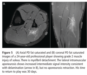
Grade 2 injury to soleus. Source: Prakash et al., (2018)
The calf muscles are comprised of 3 muscles – the medial gastrocnemius, lateral gastrocnemius and the soleus. Each muscle has a layer of a flattened sheet of tendon tissue called aponeurosis. The soleus muscle has a more complex connective tissue structure as it also has an intramuscular aponeurosis which separates its anterior compartment from the posterior compartment, and a central tendon that splits the anterior compartment(5).
The calf muscles are designed for high force generation and efficiency due to their small pennate fibres and large physiological cross-sectional area. This fibre arrangement is suited to isometric contractions where there are minimal length changes during a sprint or steady speed running(6). During high velocities muscle contractions such as acceleration or jumping, there is some fast shortening and rotation of the muscle fibres to apply a high rate of force to the Achilles tendon for an amplified energy return(6,7). The soleus muscle is the largest force contributor of the calf muscle complex(8) and is predominantly slow-twitch fibre composition which facilitates high work rates over sustained periods. It contributes 50% of vertical support during the stance phase of running and change of direction (9,10).
Achilles Injuries
Achilles tendinopathy is an overuse injury that can occur at the mid-portion of the tendon or at the insertion to the heel. It has an incidence rate of 7-9% in running-based sports and around 50% of athletes will experience this injury during their lifetime(11). Athletes who develop this injury typically present with reduced calf strength and hop performance, and often report a big increase in intensity in the weeks prior to onset(12).
Achilles tendon ruptures occur when the ankle goes into rapid dorsiflexion during ballistic movements such as accelerating and jumping, where the tendon undergoes a fast stretch with delayed recruitment of the calf muscles(13). Tendon tissue quality may be a factor as these injuries can occur during pre-season after a long period of inactivity. The most famous incidence of these injuries was in the period after the NFL lockout in 2011 where a cluster of Achilles ruptures were reported in the period of return to training. There is also some evidence of pathological changes occurring within the tendon prior to a rupture(14). In a cohort of NFL players who suffered an Achilles rupture, 61% successfully returned to sport, meaning that around 40% of athletes who fail to return to the same level of performance(15). A rupture and surgical repair can lead to impairments in muscle-tendon unit function and lower limb biomechanics. This is due to loss of tension in the tendon and reduced fascicle length of the calf muscles leading to a reduction ankle joint power(16).
During high-speed running and maximal effort hopping the Achilles tendon undergoes tensile forces of over 10 times body weight(17). Tendon stiffness describes the ability to resist a stretch under force. A tendon with an optimal level of stiffness can store more elastic energy to be reutilised during propulsion. Strains between 4-8% are required in order for the tendon to improve its stiffness but the force required to achieve those strains can vary from person to person(18). Tendons are mechanically sensitive and metabolically active organs. The cells that reside between the collagen fibres react when they sense a certain level of strain and force. If the tendon doesn’t get exposed to these strains and tensile forces on a regular basis, it deconditions and it’s stiffness properties can reduce(19). Conversely, acute exposure to repetitive levels of high strains with insufficient recovery can lead to a maladaptive response and increased risk of injury(19).
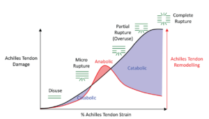
Optimal Achilles tendon strain for an adaptive response. Source: Pizzolato et al., (2019)
Calf and Achilles Assessments
It is useful to assess calf strength and hop performance to profile an athlete’s muscle-tendon unit qualities. A measure of isometric strength relative to body weight can be done with force plates if available or using a leg press or smith machine. When using force plates with an isometric rig, we can test with a straight knee to get a measure from all the calf muscles. For uninjured or fully rehabilitated elite athletes, we see typical peak force values above 3 times bodyweight. However, depending on the setup and cueing, it can be difficult to control contribution from the knee and hip extensors to vertical force achieved. We can also perform the test seated with the knee at a 90° angle. This narrows down the force contribution to the soleus muscle as it is operating on a favourable region of its force-length curve, while the gastrocnemius muscles are too short to contribute sufficient force(20). Force values of more than twice bodyweight is a widely accepted return to sport or pre-season outcome measure in field sports players.
Testing Achilles tendon stiffness during isometric contractions require an isokinetic dynamometer, ultrasound, 3D motion capture, and a skilled clinician to perform the measurements. This is not usually feasible in performance or clinical settings. We can get a surrogate measure of tendon qualities using the hop test. Achilles tendon stiffness has a strong relationship with ground contact time and jump height in a drop jump, as well as hop distance in a horizontal hop(21,22). A horizontal hop can also provide a useful measure of soleus function given that it has been shown to provide the largest muscle contribution to the propulsion phase(23).
Training the Calf and Achilles
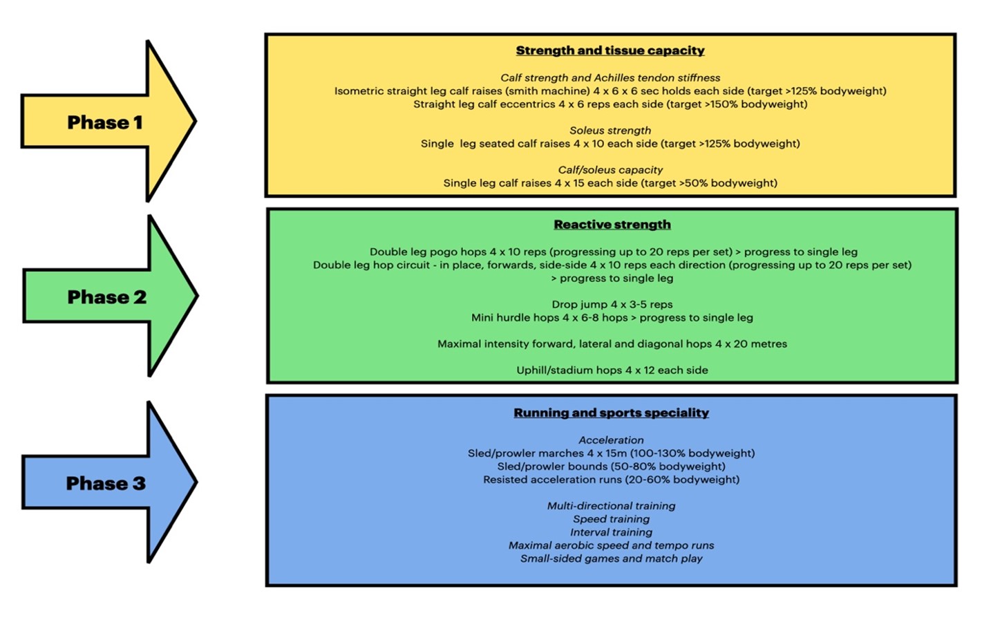
A layered approach is advised to prepare the calf complex for a return to competitive sport. The initial focus would be on developing strength and tissue capacity with heavy-loaded resistance exercises. We can select exercises to be more specific with muscle and tendon adaptations. Heavy isometric or concentric training favours increases in pennation angle and force capacity(24). Heavy eccentric training leads to increases in fascicle length along with tendon stiffness and connective tissue remodelling(25,26). Isometric exercises can be performed to increase force capability at certain joint angles or muscle-tendon unit lengths. It is also important to consider training calf endurance qualities depending on the athlete and the sport involved.
The next phase is perhaps the most critical – developing reactive strength qualities. The muscle and tendon forces and loading rates closely replicated the demands of high-speed running(27). Depending on the profile of the athlete you can work both ends of the spectrum with this – low volume high-intensity plyometrics and high volume moderate intensity plyometrics. Horizontal hopping and incline hopping will greatly challenge the contractile and connective tissue structures of the calf complex.
Most calf and Achilles injuries occur as running intensities and volumes increase. It is important that this is periodised appropriately with a gradual progression to the high-intensity running demands of the sport. The recently published “Return to Training Guidelines” for GAA and “Competition Ready Guidelines” for World Rugby are highly recommended resources for preparing for running and match-play demands for field sports. An example of a more specific progression pathway for calf and Achilles is outlined below.
Setanta College has been delighted to partner with the GAA to deliver the ‘Be Ready to Play’ programme, which aims to help players and coaches navigate the return to play safely, and you can access it here.
References
- Prakash A, Entwisle T, Schneider M, Brukner P, Connell D. Connective tissue injury in calf muscle tears and return to play : MRI correlation. 2018;929–33.
- Mackey AL, Kjaer M. REVIEW Recovery from Exercise Connective tissue regeneration in skeletal muscle after eccentric contraction-induced injury. J Appl Physiol [Internet]. 2017 [cited 2020 Oct 20];122:533–40. Available from: http://www.jappl.org
- Green B, Lin M, McClelland JA, Semciw AI, Schache AG, Rotstein AH, et al. Return to Play and Recurrence After Calf Muscle Strain Injuries in Elite Australian Football Players. Am J Sports Med [Internet]. 2020 Oct 8 [cited 2020 Oct 11];036354652095932. Available from: http://journals.sagepub.com/doi/10.1177/0363546520959327
- Pedret C, Rodas G, Balius R, Capdevila L, Bossy M, Vernooij RWM, et al. Return to Play After Soleus Muscle Injuries. Orthop J Sport Med. 2015;3(7).
- Hodgson JA, Finni T, Lai AKM, Edgerton VR, Sinha S. Influence of Structure on the Tissue Dynamics of the Human Soleus Muscle Observed in MRI Studies During Isometric Contractions. J Morphol. 2007;268(February):254–74.
- Lai A, Brown N, Lai A, Schache AG, Brown NAT, Pandy MG, et al. Human ankle plantar flexor muscle – tendon mechanics and energetics during maximum acceleration sprinting Human ankle plantar flexor muscle – tendon mechanics and energetics during maximum acceleration sprinting Author for correspondence : 2016;(August).
- Roberts TJ, Eng CM, Sleboda DA, Holt NC, Brainerd EL, Stover KK, et al. The Multi-Scale, Three-Dimensional Nature of Skeletal Muscle Contraction. Physiology.2019;34(6):402–8.
- Roy RR, Shellock FG, Hodgson JA, Edgerton VR, Fukunaga T. Specific tension of human plantar flexors and dorsiflexors. J Appl Physiol. 2017;80(1):158–65.
- Dorn TW, Schache AG, Pandy MG. Muscular strategy shift in human running: dependence of running speed on hip and ankle muscle performance. J Exp Biol [Internet]. 2012;215(13):2347–2347. Available from: http://jeb.biologists.org/cgi/doi/10.1242/jeb.075051
- Maniar N, Schache AG, Cole MH, Opar DA. Lower-limb muscle function during sidestep cutting. J Biomech. 2019 Jan 3;82:186–92.
- Kujala UM, Sarna S, Kaprio J. Cumulative incidence of achilles tendon rupture and tendinopathy in male former elite athletes. Clin J Sport Med. 2005;15(3):133–5.
- Järvinen TAH, Kannus P, Maffulli N, Khan KM. Achilles tendon disorders: Etiology and epidemiology. Foot Ankle Clin. 2005;10(2):255–66.
- De la Fuente C, Ramirez-Campillo R, Gallardo-Fuentes F, Alvarez C, Bustamante C, Henríquez H, et al. Pattern analysis of a complete Achilles tendon rupture suffered during high jump preparation in an official national-level athletic competition. Sport Biomech [Internet]. 2019;00(00):1–11. Available from: https://doi.org/10.1080/14763141.2019.1651897
- Maffulli N, Via AG, Oliva F. Chronic Achilles Tendon Disorders. Clin Sports Med [Internet]. 2015 Oct;34(4):607–24. Available from: https://linkinghub.elsevier.com/retrieve/pii/S0278591915000617
- Yang J, Hodax JD, Machan JT, Krill MK, Lemme NJ, Durand WM, et al. Factors Affecting Return to Play After Primary Achilles Tendon Tear: A Cohort of NFL Players. Orthop J Sport Med [Internet]. 2019 Mar 12;7(3):232596711983013. Available from: http://journals.sagepub.com/doi/10.1177/2325967119830139
- Baxter JR, Farber DC, Hast MW. Plantarflexor fiber and tendon slack length are strong determinates of simulated single-leg heel raise height. J Biomech [Internet]. 2019;(xxxx):1–7. Available from: https://doi.org/10.1016/j.jbiomech.2019.01.035
- Komi P V. Relevance of in vivo force measurements to human biomechanics. J Biomech. 1990;23(SUPPL. 1).
- Arampatzis A, Mersmann F, Bohm S. Individualized Muscle-Tendon Assessment and Training. Front Physiol [Internet]. 2020 Jun 26 [cited 2020 Jul 26];11:723. Available from: https://www.frontiersin.org/article/10.3389/fphys.2020.00723/full
- Pizzolato C, Lloyd DG, Zheng MH, Besier TF, Shim VB, Obst SJ, et al. Finding the sweet spot via personalised Achilles tendon training: The future is within reach. Br J Sports Med. 2019;53(1):11–2.
- Arampatzis A, Karamanidis K, Stafilidis S, Morey-Klapsing G, DeMonte G, Brüggemann GP. Effect of different ankle- and knee-joint positions on gastrocnemius medialis fascicle length and EMG activity during isometric plantar flexion. J Biomech. 2006;39(10):1891–902.
- Abdelsattar M, Konrad A, Tilp M. Relationship between achilles tendon stiffness and ground contact time during drop jumps. J Sport Sci Med. 2018;17(2):223–8.
- Wang HK, Lin KH, Su SC, Shih TTF, Huang YC. Effects of tendon viscoelasticity in Achilles tendinosis on explosive performance and clinical severity in athletes. Scand J Med Sci Sport. 2012;22(6):1–9.
- Kotsifaki A, Korakakis V, Graham-smith P, Sideris V, Whiteley R. Vertical and Horizontal Hop Performance : 2021;XX(X):1–8.
- Franchi M V, Reeves ND, Narici M V, Huey K. Skeletal Muscle Remodeling in Response to Eccentric vs . Concentric Loading : Morphological , Molecular , and Metabolic Adaptations. 2017;8(July):1–16.
- Geremia JM, Manfredini B, Maarten B, Bobbert F, Rico R, Juner F, et al. Effects of high loading by eccentric triceps surae training on Achilles tendon properties in humans. Eur J Appl Physiol [Internet]. 2018;0(0):0. Available from: http://link.springer.com/10.1007/s00421-018-3904-1
- Geremia JM, Baroni BM, Bini RR, Lanferdini FJ, de Lima AR, Herzog W, et al. Triceps Surae Muscle Architecture Adaptations to Eccentric Training. Front Physiol [Internet]. 2019 Nov 26;10. Available from: https://www.frontiersin.org/article/10.3389/fphys.2019.01456/full
- Baxter JR, Corrigan P, Hullfish TJ, O’Rourke P, Silbernagel KG. Exercise Progression to Incrementally Load the Achilles Tendon. Med Sci Sports Exerc. 2021;53(1):124–30.
Tendinosis not tendinitis – the physician and sports medicine – pub med – 2000
Points up the damage to tendons: is the result of mechanical strain, and overuse (RSI) in which the muscle spindle cells are over active, affecting a chronic tension ( 24/7 ) that defeats the tendon’s re-modeling of the collagen that has been damaged and thus risks further damage and possible partial or full tear.
When the muscle body is contracted – and not capable of fully relaxing, the lack of re-modeling of collagen will continue to be at risk. The most effective approach is to overstimulate the involved tissue ( tendon-muscle body- tendon ) by high pressure deep tissue massage and cause the overactive muscle spindle cells to reduce their ability to contract the muscle tissue. Over daily massage this stripping massage will give space to return the muscle group to normal relaxed length – which gives collagen a lessened loading and the “space” to remodel properly.
Pain of the tissue to low levels of massage pressure indicates muscle overuse injury. The translation from German muscle pain research in the early 1900’s of the article by Dr. David Simons is instructive.